-
 Melatonin Research
Vol 7 No 3 (2024)
Melatonin Research
Vol 7 No 3 (2024)While all the articles in this issue are interesting, three seem especially timely. In an in silico study, Acharyya and Hasan systemically studied the potential therapeutic effects of melatonin on COVID-19 and compared its pharmacokinetics with clinically commonly used therapeutic medicines against COVID-19 including methylprednisolone, doxycycline, oseltamivir, and remdesivir. The results showed that melatonin had obvious advantages over these medicines in the properties of absorption, distribution, metabolism, and excretion (ADME). In addition, molecular docking study indicated the substantial interactions of melatonin with principal hub targets of COVID-19 including TP53, AKT1, IL6, TNF, IL1B, BCL2, EGFR, STAT3, CASP3, and NFKB1. These molecules are also involved in the pathologies of many other diseases. The protein-to-protein interaction (PPI) analysis suggested that melatonin may directly bind to these proteins with hydrogen binds to stimulate or inhibit their actions depending on the configuration. These results provide novel insights for understanding the multifaceted actions of melatonin used in many different disorders. In another study, Valiensi et al reported a cross-sectional study on the use of high doses of melatonin in humans. The significantly high doses of melatonin between 40 and 200 mg per day, with a mean of 76.56 mg ± 33.58 mg daily were given to patients over 4 months. These amounts of melatonin did not induce noticeable side effects or to interfere the concomitant pharmacological treatment. The results confirm the safe use of melatonin at these dose levels. This is especially relevant for some devastating diseases which currently have no effective treatments such as COVID-19. A third study relates to the origin of melatonin synthetic enzyme, serotonin N-acetyltransferase (SNAT). SNAT has been identified in eukaryotes, bacteria, i.e., two of the three domains of life. Its present in archaea was only a prediction. In a current study, Lee, and Back cloned an archaeal SNAT from Thermoplasma volcanium (TvSNAT) and overexpressed it in rice lines. These TvSNAT-OE lines showed high melatonin levels with delayed senescence, but were more susceptible to salt stress than the WT due to melatonin induced enhanced brassinosteroid synthesis. The results further support that SNAT is present in all three domains of life as is melatonin.
-
 Melatonin Research
Vol 7 No 2 (2024)
Melatonin Research
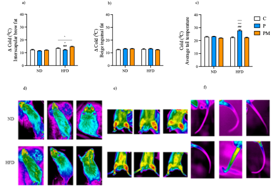
Vol 7 No 2 (2024)In the current issue, Chacín-Bonilla and Bonilla have thoroughly reviewed the potential biological actions of melatonin in reducing viral pathophysiology. They claimed that as a potent antioxidant, anti-inflammatory agent, a stimulator of immune functions, and regulator of apoptosis, melatonin is suitable a relative for use in viral infections, which are often associated with excessive inflammatory responses and elevated oxidative stress. In addition, the virus- and cytokine- storm-driven control of the pineal and mitochondrial melatonergic pathway to disrupt the immune responses and increase gut dysbiosis, suppressing levels of the short-chain fatty acid, butyrate, and increasing circulating lipopolysaccharides, stimulating viral replication and host symptoms severity, melatonin supplementation can reverse these pathological alterations. Additional interesting observations in this issue are reported by Belpiede et al. They observed transgenerational effects of that the loss of the circadian melatonin rhythm of the mother altered energy metabolism of their offspring. Their results showed that maternal pineal melatonin rhythm loss due to pinealectomy disrupted the energy metabolism of the offspring while melatonin replacement of the pinealectomized mother normalized the energy metabolism in the offspring submitted to the high-fat diet, enabling them to make functional adaptations such as the reduced food consumption, greater thermoregulatory capacity as well as reduction of white adipose tissue mass. These transgenerational effects of maternal melatonin level have rarely been reported. Another important and relevant observation reported in this issue is that melatonin exhibits different effects on adipose tissues under normoestrogenic and estrogen-deficient conditions in rats. Hermoso et al observed that in ovariectomized (OVX) rats, melatonin treatment suppressed the visceral retroperitoneal adipose tissue and increased the prevalence of small adipocytes of subcutaneous inguinal adipose tissue, suggesting a better lipid distribution among adipose tissues to minimize the risks for the development of metabolic abnormalities due to estrogen deficiency. However, under normoestrogenic condition, melatonin reduced plasma estradiol levels and uterine mass, raising concerns about its effect on reproductive functions. These unexpected observations require confirmation.
-
 Melatonin Research
Vol 7 No 1 (2024)
Melatonin Research
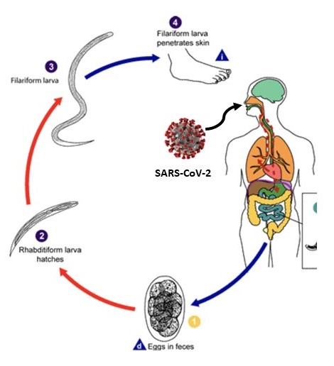
Vol 7 No 1 (2024)In the current issue, Leonor and Ernesto Bonilla suggest and provide the evidence that supports the likely therapeutical utility of melatonin for the co-infection of SARS-CoV-2 and intestinal helminths. The immunoregulatory role of intestinal helminths influence the progression of SARS-CoV-2 infection and vaccination effectiveness. A preexisting helminth infection could impair the host’s ability to fight off SARS-CoV-2 and augment morbidity and fatality. For example, the load of soil-transmitted helminth infections among the Amerindians of the Brazilian Amazon was high and the mortality rate of COVID-19 in these regions was 250% higher than that in the rest of the country. If helminthiases increase complications, then the burden of COVID-19 in helminth endemic countries might be worse than expected. Since melatonin has beneficial effects on both viral and parasitic infections and it is economically affordable in most of the developing countries and regions, its use for this co-infection may be an optimal choice and should be considered. A second interesting study published in this issue is that of Hernando et al. The authors examined prolonged photoperiodic alterations (15 days constant light exposure or 15 days constant dark) plus iron-overloading in rats in reference to their serum melatonin levels and the amount of brain lipid peroxidation. The results showed that the reduced melatonin level caused by photoperiodic changes and iron-overloading is negatively associated with the brain lipid breakdown. However, the unexpected observation is that when the rats initially in the prolonged photoperiodic alterations were returned to a normal light/dark cycle for 15 days, they still exhibited an abnormal melatonin pattern compared to the control. This interesting and preliminary observation requires further confirmation by others.
-
 Melatonin Research
Vol 6 No 4 (2023)
Melatonin Research
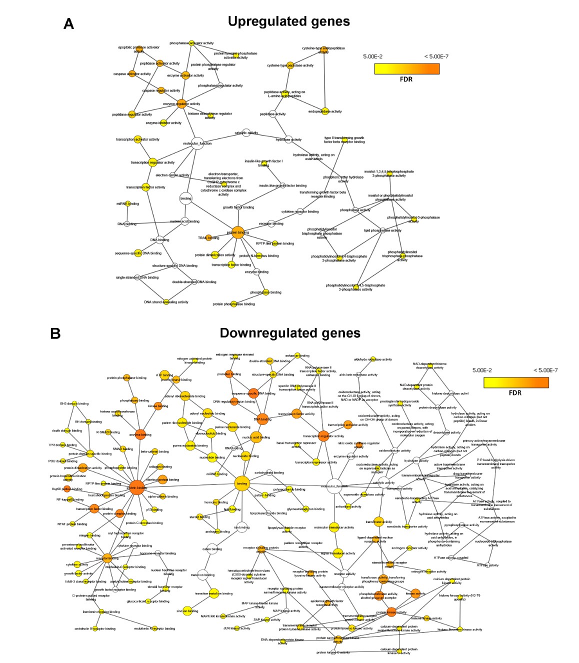
Vol 6 No 4 (2023)The anti-cancer action of melatonin has been extensively studied in cell culture, animal models or clinical research from the different aspects. In the current issue, a publication by Chuffa et al. summaries these results and examines some of the mechanisms involved. To help to clarify the molecular processes by which melatonin modulates carcinogenesis, the authors for the first time explore the cancer-melatonin interactions at the gene network level with the use of text-mining strategies. The results indicate a close relationship among breast, hepatocellular, prostate, and oral cancers, as well as neuroblastoma and osteosarcoma in terms of the melatonin-related signaling pathways. Melatonin upregulated genes promote apoptotic protease activator, caspase activator, enzyme regulator, and protein binding, whereas the downregulated genes influence protein kinase activities, transcription factor binding, numerous proteins and enzymes, DNA, and promoter bindings in cancer cells. Clinically, melatonin-downregulated genes are associated with the longer survival of patients with glioblastoma, bladder, breast, colon, stomach, liver, lung, and ovarian carcinomas. These results provide a global view of gene interaction networks in melatonin-treated cancers and their functional value, opening new opportunities to consider melatonin for cancer therapy. In another report, Santillán-Morales and Benítez-King present a novel idea to use olfactory neural precursors (ONPs) as a model that reflects biological mechanisms of neurons from CNS to study the Alzheimer’s pathologies and treatment effects of melatonin. While it is generally impractical to obtain life neurons from the CNS of patients with Alzheimer’s disease (AD), it is readily feasible to collect the ONPs from patients’ nostril noninvasively. The easy availability of live ONPs which have the characteristics of potential neurogenesis from the AD patients provides a useful new tool in which to test melatonin effects on this debilitating disease.
-
 Melatonin Research
Vol 6 No 3 (2023)
Melatonin Research
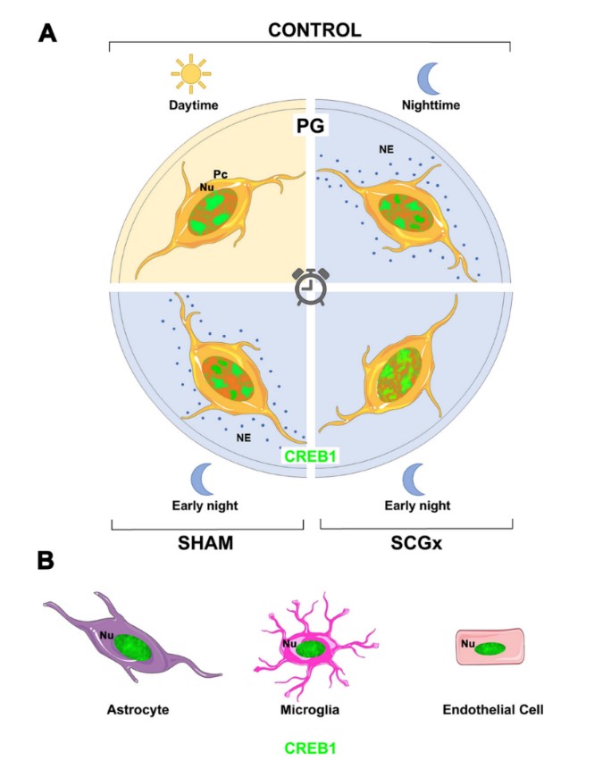
Vol 6 No 3 (2023)In the current issue, two studies have addressed the potential regulatory mechanisms of melatonin biosynthesis in remotely related species. One is in the pineal gland of an animal (rat) and another is in the fruit of a plant (pepper). It is obvious that they utilize different regulatory pathways but both use environmental light as the clue to adjust the transcriptional activities related to circadian rhythmicity and possibly for melatonin production. This is a conserved process that involves a large range of species. As reported by Farias Altamirano et al., in pineal gland, the major driver of the circadian rhythmicity seems to be CREB1. The study found that the overall transcription activity of CREB1 is mostly conserved between the light and dark phases; however, a daily dynamism of the nuclear domains occupied by CREB1 is present only in pinealocytes, with larger dispersion during the dark phase when the pinealocytes are under the influence of norepinephrine (NE) released from postganglionic sympathetic neurons. The high concentration of nuclear CREB1 in non-pinealocyte cells including astrocytes, phagocytes and endothelial cells was not observed. This indicates CREB1 participates in the nocturnal melatonin synthesis that rhythmically modulates the physiology and behavior of the organism. In plants, particularly in the pepper plant (Capsicum annuum L.), Taboada et al. have for the first time identified several tryptophan decarboxylase (TDCs) genes which are the initial enzymes for melatonin biosynthesis in plants. Of the two novel TDCs found in the pepper fruit, CaTDC4 is positively modulated by two light-responsive elements, Box4 and TCT-motif, and CaTDC5 is regulated by GT1-motif and G-Box. This is in good agreement with the mechanism of regulation of melatonin biosynthesis by different light conditions including the presence/absence of light and the intensity of light on circadian rhythms. Interestingly, these two TDCs are differentially expressed, i.e., when CaTDC4 is upregulated, CaTDC5 is downregulated. As is known, the regulatory mechanisms of melatonin biosynthesis in plants are more complex than those in animals. How these mechanisms evolved from complex to simple from low-rank life forms to high-rank organisms is currently unknown, but this issue will surely be resolved as enthusiasm for this research continues to attract scientists to the field.
-
 Melatonin Research
Vol 6 No 2 (2023)
Melatonin Research
Vol 6 No 2 (2023)Several papers related to novel aspects of melatonin physiology are published in this issue. One is the potential association of melatonin and Helicobacter pylori (H. pylori) infection. H. pylori infection is the culprit in several disorders of the gastrointestinal tract, including gastric cancer. H. pylori infection reduces melatonin synthesis in the gastric epithelial cells while melatonin supplementation ameliorates gastric disorders. The beneficial effects of melatonin on H. pylori infection may involve the antioxidative, anti-inflammatory activities of melatonin as well as its regulation on DNA damage response and epigenetic modifications. These features make melatonin a suitable candidate as an adjuvant for H. pylori infection treatment. Another interesting observation published in this issue relates to how melatonin penetrates the cell membrane. One classic opinion is that due to the high lipophilic nature of this molecule, melatonin freely enters the cell by diffusion. With the use of a 3D cell culture model, Mayo et al identified that only a small portion of melatonin enters a cell by diffusion. Thus, the active uptake via common or specific transporters of this molecule seems to be responsible for its intracellular accumulation. This observation may be applicable as how to efficiently improve the intracellular melatonin levels especially, for example, when it is used as a cancer treatment. In addition, Miki et al report that melatonin is more effective on bone metabolism when given at early night than during the day in ovariectomized rats. Even though this observation is contrary to some previously published data, it seems consistent with melatonin’s circadian rhythm in the blood.
-
 Melatonin Research
Vol 6 No 1 (2023)
Melatonin Research
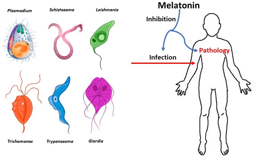
Vol 6 No 1 (2023)In the current issue, Cárdenas et al. have introduced a new strategy for the use of melatonin to control the tropical parasitic infections including Plasmodium, Toxoplasma, Trypanosoma, Leishmania, Schistosoma and others. The focus is on the antioxidant, anti-inflammatory and immunoregulatory activities of melatonin on the parasitic invasions and their related pathologies. Especially, in malaria, melatonin seems to synchronize the Plasmodium life cycle with the host circadian changes. In animal studies, disrupting its life cycle with melatonin intervention can control the severity of Plasmodium infection. The Trypanosoma infection evolves into chronic Chagas disease, representing a public health problem with grave repercussions in Latin America and other areas of the world. Melatonin treatment augments the levels of TNF-α, IFN-γ, and IL-12 of the host to protect against this parasitic infection. Parasitic diseases still represent a significant burden on health systems worldwide, particularly in low and lower-middle income countries with limited access to sanitation facilities and resources for therapeutic approaches. However, the continuing study of melatonin, as readily available, affordable and fundamentally safe healing option, might help better control these infections.
-
 Melatonin Research
Vol 5 No 3 (2022)
Melatonin Research
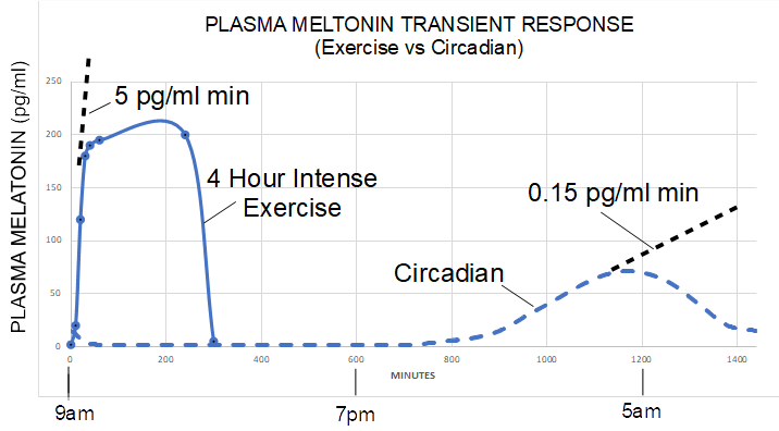
Vol 5 No 3 (2022)One of the most interesting and thought-stimulating papers in the current issue is the report published by Skutsch et al.; they noted that COVID-19 deaths per million were higher in South America than in either Europe and North America, while Asia, Africa, and Oceania had death rates which were only a fifth of those in South America. They found that the COVID-19 death rate was strongly associated with overweight and high latitudes but not with the vaccine coverage percentage in these countries. In an attempt to explain these differences, they hypothesized that: (1) In overweight people there is less penetration of near-infrared radiation (NIR) to the depth of important organs; stimulation of these organs by NIR would result in elevated production of subcellular melatonin, a strong antioxidizing factor. (2) In overweight people, fatty tissue holds much of the body´s 25(OH)D3 leaving less circulating in the blood making it less systemically protective. The hypothesis advanced by Skutsch et al. receives support from an article by Zimmerman and Reiter published also in this issue. They observed that large quantities of melatonin, greater than 5 pg/ml min ramp rates for plasma and sweat melatonin in human subjects, have been detected during strenuous exercise in sunlight as compared to 0.15 pg/ml min ramp rates for plasma melatonin under dim light melatonin onset conditions. This difference is in excessive of 30-fold. Sunlight contains high levels of NIR which likely stimulate mitochondrial melatonin production. High latitude has less NIR irradiation and excessive weight restricts NIR penetration to the important organs such as the lungs and heart. Thus, both obesity and high latitude are factors that limit local melatonin production and compromise the protective effects of locally-produced melatonin in these important organs. These observations may not only apply to COVID-19 patients but to other disorders including diabetes, neurodegenerative diseases and seasonal depression.
-
 Melatonin Research
Vol 5 No 2 (2022)
Melatonin Research
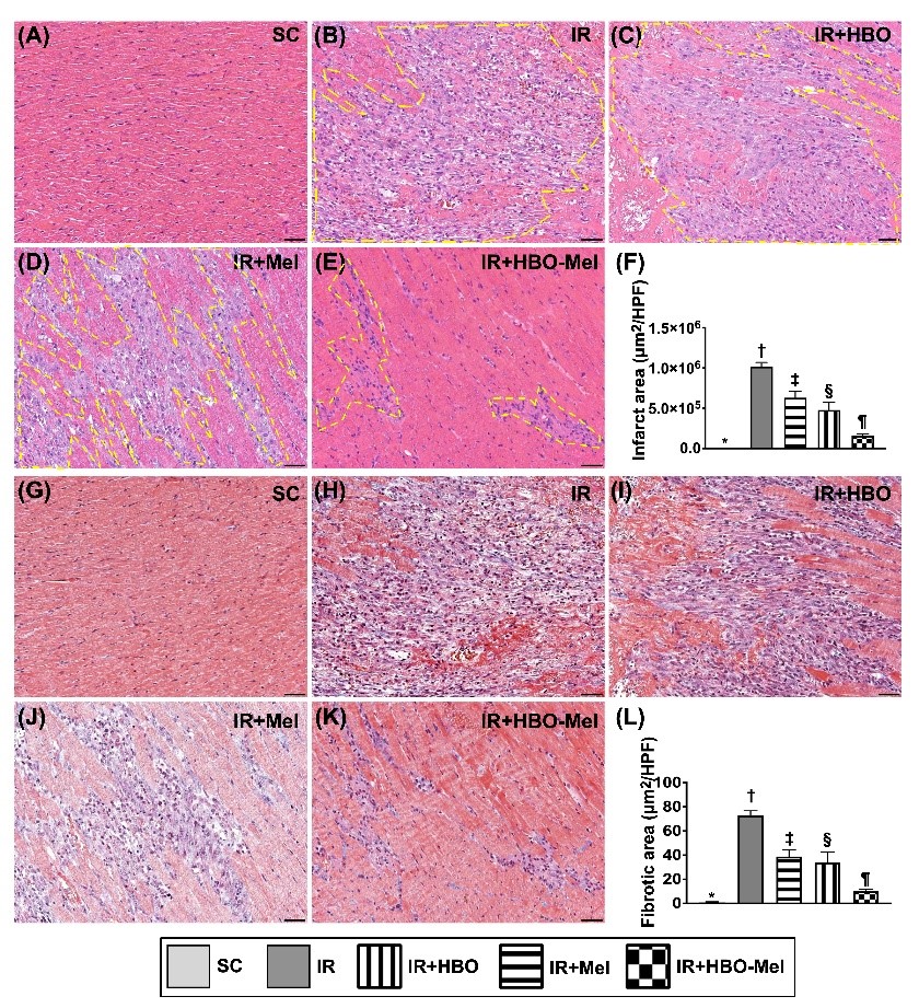
Vol 5 No 2 (2022)Two important observations related to melatonin’s potential therapeutic effects are uncovered in this issue. Chai et al. using both an in vitro and in vivo model documented, for the first time, that melatonin combined with hyperbaric oxygen therapy is an efficient means to shield the heart from acute ischemia-reperfusion injury. The evidence shows that this combination protected against the acute phase of ischemia-reperfusion cardiac damage including cardiac infarct size and ejection function of the heart. Moreover, the combined therapies also improved the post-ischemia-reperfusion cardiac remodeling by inhibiting the cardiac fibrosis; this latter observation is supported by earlier published findings. These observations obviously have high clinical relevance and should encourage clinical trials using this therapy. In another clinically significant observation, Wongchitrat et al. are the first to report that melatonin has suppressive actions on Zika virus (ZIKV) replication in different cell lines including human neural cells. One molecular mechanism involves melatonin’s high binding affinity to the ZIKV non-structural 3 (NS3) protein which is a necessary component allowing the ZIKV to enter cells. By interfering with the entrance of the ZIKV into cells, melatonin reduces its infectivity. Currently, there are no effective antiviral medications available to treat ZIKV infections; thus, melatonin may provide a means to effectively prevent and/or treat ZIKV-mediated pathologies. This observation warrants further investigations using animal models with the intent of extending them to human trials. Clinical trials using melatonin are readily feasible because of its high safety profile over a wide range of doses, its low cost, and the virtual absence of serious side effects.
-
 Melatonin Research
Vol 5 No 1 (2022)
Melatonin Research
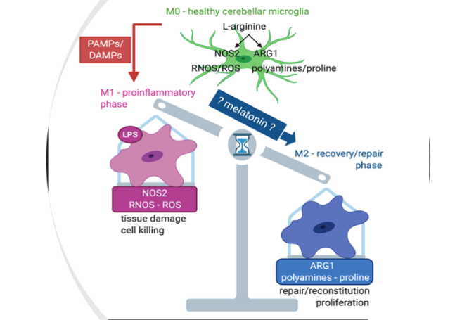
Vol 5 No 1 (2022)In this issue, a wide range of interesting topics is covered. These include documentation of the bacteriostatic properties of melatonin, the effects of melatonin on parasite development inside the host, the protective effects of a combination of melatonin with paclitaxel on ovarian carcinoma cells and the effects of pineal melatonin on the mRNA expression of melatonin and steroidogenic-related receptor genes in the rat reproductive system, etc. We also want to recommend two important research articles that appear in the current issue. One is that of Souza et al. and other is reported by Ko et al. Souza et al. show that melatonin synthesized by microglia plays a key role in the transformation of microglia type 1 (M1) to microglia type 2 (M2) phenotypes. M1 is responsible for tissue pro-inflammatory reactions and in many cases, it causes the severe tissue damage observed such as in the SARS-CoV2 infection while M2 produce anti-inflammatory actions to cope with injury recovery. This also indicates melatonin administration will help the M1 to M2 transition and suppress the inflammatory tissue injury in many different situations. Ko et al. report that by using 31phosphorus-magnetic resonance spectroscopy (31P-MRS), they can directly measure parameters of liver metabolic function in the intact animals and thus, this method can monitor the detailed evolution of liver fibrosis. To the best of our knowledge, this is the first use of 31P-MRS for this purpose. In addition, with the aid of this method they observed that when melatonin-pretreated mitochondria are transfused into the blood of the liver-damaged animals, the hepatic fibrosis of the animals is significantly reduced. The results provide a new avenue to target the pathophysiology of the liver fibrosis which currently lacks an effective treatment. We hope that the novel observations reported in the current issue will stimulate the enthusiasm of melatonin scientists to further advance this important field by applying melatonin to other new experimental and clinical situations.
-
 Melatonin Research
Vol 4 No 4 (2021)
Melatonin Research
Vol 4 No 4 (2021)In this issue, the potential utility of melatonin to improve rice production during the ongoing global climate change is discussed. It has been estimated that the global annual rice production is around 782 million tons and it contributes to the food supply of more than half of the world’s population. However, because of the global warming, the annual yield of rice will be dramatically reduced since this grain is sensitive to the elevated ambient temperature. To secure the food supply for the continuously-increasing population, scientists must find a way to, at least, maintain rice production at the highest level. A feasible strategy has been proposed by Back et al. They suggest the manipulation of melatonin enriched-rice strains using transgenic technology, i.e., generating rice strains that overexpress melatonin synthetic genes SNAT or ASMT. Elevated levels of melatonin would enhance the tolerance of rice against abiotic stresses resulting from heat and draught and would maintain rice production even under hot environments. Several studies have provided preliminary results to support this strategy. Another important topic considered in the current issue is the recent report in which melatonin treatment dramatically reduced the mortality of the severe COVID-19 patients. The COVID-19 pandemic has persisted for two years with no signs of abatement primarily due to the continued emergence of new variants. These new variants persistently challenge the efficacy of the vaccines. This makes the development of antiviral drugs urgent but difficult. Pharmaceutical companies reportedly have now developed drugs for treatment of COVID-19 patients. However, some of these medications cost thousand dollars per a course of treatment. Patients in many developing countries cannot afford these expensive therapies. Additionally, the majority of these drugs are designed to treat mild to moderately severe patients. Hence, there is no effective treatment for the severe COVID-19 cases, especially for the purpose of lowering their mortality. Recently, Hasan et al. reported that conventional treatment plus melatonin reduced mortality rate by 93% in severely-ill COVID-19 patients compared to the conventional treatment alone. The cost of melatonin for a course of treatment is about 5 US dollars. It is thus affordable by essentially all patients in every country; this is in addition to its high safety profile and its ease of oral use. If the observations reported by Hasan and colleagues are confirmed, melatonin should be recommended for treatment of the severe COVID-19 patients, especially in the underdeveloped countries where conventional health care is not readily available. Also in this issue, Bitar et al. proposed a novel association between melatonin and neurodegenerative diseases. They believe that the neural glymphatic system is the highway to transport metabolic by-products and debris from the brain parenchyma and subarachnoid cerebrospinal fluid (CSF) with this well-functioning network possibly slowing the onset of neurodegenerative diseases. The clearance of amyloid-β reportedly occurs especially during slow wave sleep which happens concurrently with highest glymphatic drainage and with the maximal CSF levels of melatonin. Melatonin administration defers amyloid-β buildup in the brain of animals and causes its accumulation in the cervical lymph nodes. With increased age CSF melatonin levels drop markedly, co-incident with neurodegeneration and dementia. These findings suggest a potential association between the loss of melatonin, decreased glymphatic drainage and neurocognitive decline in the elderly.
-
 Melatonin Research
Vol 4 No 3 (2021)
Melatonin Research
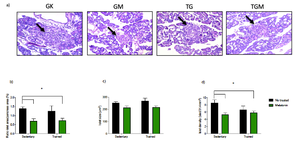
Vol 4 No 3 (2021)In this issue, Leite et al. report that a combination of melatonin and moderate-intensity aerobic exercise improves pancreatic beta-cell function and glycemic homeostasis. This study was performed using the Goto-Kakizaki (GK) rats which frequently serve as the type 2 diabetic animal model due to their spontaneously-occurring hyperglycemia and the elevated resistance of peripheral tissues to insulin. In this model, melatonin treatment alone reduced the mass of epididymal white adipose tissue (WAT)); however, a combination of melatonin and physical exercise significantly reduced caloric intake, body weight, WAT and improved glucose tolerance, insulin sensitivity and also reduced apoptosis of cells in pancreatic islets. These important findings provide a potential alternative treatment for patients with type 2 diabetes since melatonin is readily available, is inexpensive and its use is essentially without side effects. Also herein, Reiter et al. suggested the continued use of melatonin to limit a COVID infection including protection against the delta variant of SARS-CoV-2 as well as any eventual future variants of the virus. Melatonin is a non-specific anti-viral agent. It not only targets the virus per se, but it also improves the host tolerance to the pathogens; thus, it has a wide spectrum of anti-viral activity including against the mutated variants. Finally, to understand the multiple biological activities of melatonin, Banerjee et al. systemically reviewed the functional developments of melatonin during evolution. They propose that the original function of melatonin served as a free radical scavenger and antioxidant for all organisms and other functions of melatonin were acquired at the different evolutionary stages. The important information in these and other articles in this issue will help to further explain the multiple beneficial effects of melatonin.
-
 Melatonin Research
Vol 4 No 2 (2021)
Melatonin Research
Vol 4 No 2 (2021)The neuroprotective effects of melatonin have been frequently studied in different pathological animal models. In some models, melatonin exhibits profound benificial effects, but in others, these actions are limited depending on the nature of the pathology and the species used. In the current issue, two reports have further investigated this issue from different perspectives. Haque et al. used cultured rat spinal cord slices and challenged them with L-glutamic acid to mimic spinal cord injury (SCI) related neuron loss. The authors observed that melatonin treatment significantly reduced cellular apoptosis and prevented penumbral neuron loss following SCI. This protective effect of melatonin was mediated by MT1/2 membrane receptors. In another study, Leung and Cheung directly injected type IV collagenase into the right striatum of rats to create intracerebral hemorrhage (ICH) pathology which is a severe form of stroke with a high mortality rate and also an important cause of permanent disability. Melatonin treatment at the dose of 50 mg/kg reduced the perihematomal microglial activation but had no effect on the size of hematoma. The microglial activation is a key factor in determing the prognosis of ICH. As a result of melatonin treatment, the neurological functions of the rats after ICH also were significantly improved. These two studies provide additional evidence that addresses the importance of melatonin in protecting against neurological pathology. We hope that these novel observations, along with those published earlier, will stimulate enthuasiam for investigations into the multipe benefits of melatonin in reducing neuropathologies including consideraton of human trials.
-
 Melatonin Research
Vol 4 No 1 (2021)
Melatonin Research
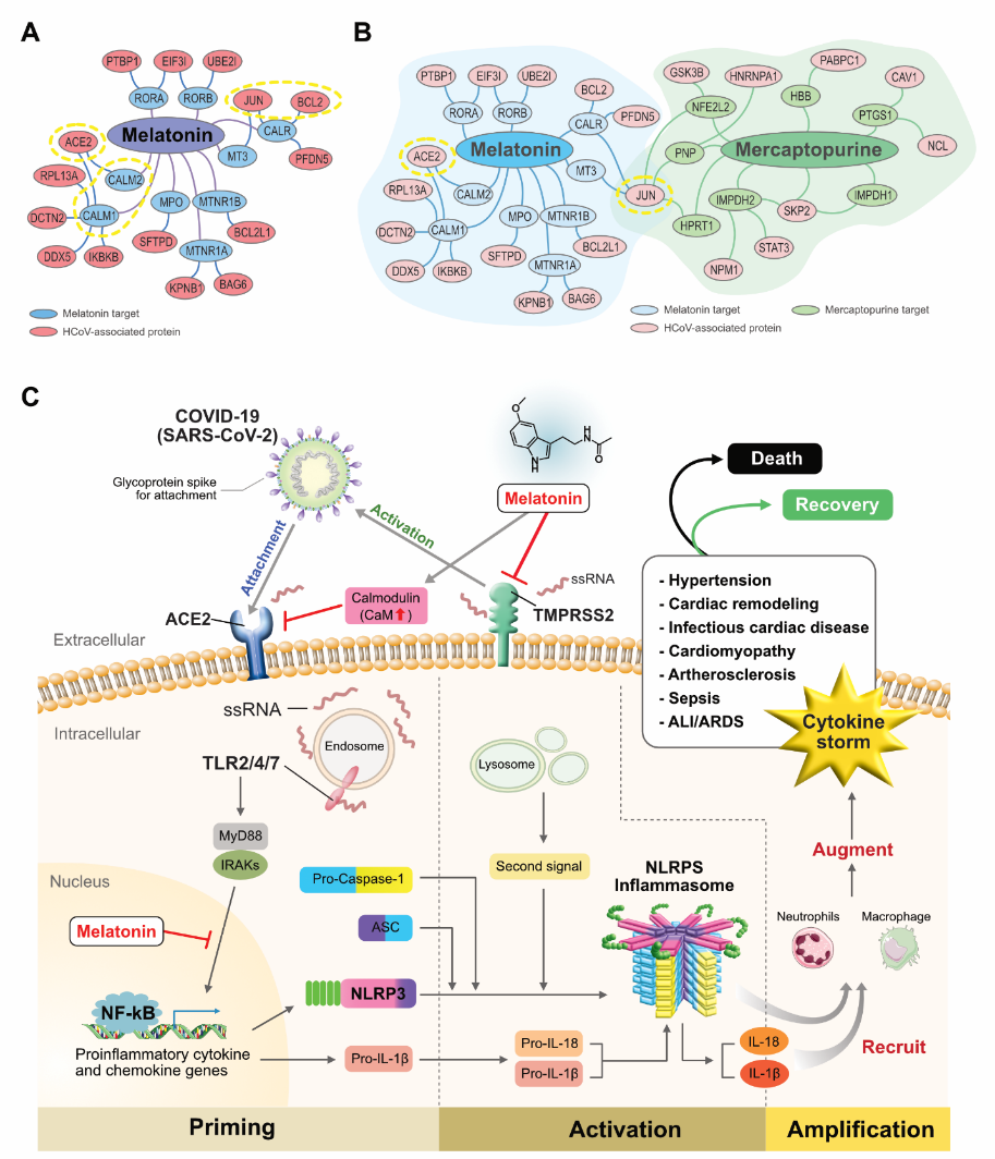
Vol 4 No 1 (2021)Although effective vaccines for SARS-CoV-2 are available in developed counties, the majority of the population in the developing world will have to wait months or even a year or more to acceess these treatments. During this relatively long period, other therapeutic methods, including melatonin, should be used to reduce the morbidity and mortality related to COVID-19. Thus, it is our intent to continously provide evidence for the potential applications of melatonin for the prevention and treatment of COVID-19 patients. In this issue, a review by Gurunathan et al. systemically introduce mechanisms as to the protective effects of melatonin on SARS-CoV-2 infection and provide comprehensive information for researchers and physiciens working in this field. Considering melatonin as a chronobiotic agent, Brusco et al. claim that properly administered, melatonin may restore the optimal circadian pattern of the sleep-wake cycle and improve clinical condition in pneumonia associated with COVID-19 patients, especially for the patients in non-intensive care unit (NICU). They provide preliminary data to show the efficacy of melatonin at the dose of 9 mg/day. In a research article by Fernandes et al, they indicate that melatonin endogenously present in the lungs is protective against COVID-19 severity by lowering the expression of genes used by SARS-CoV-2 to invade and replicate in human cells and suggest Melatonin-Index can serve as a new approach for predicting the evolution of healthy SARS-CoV-2 carriers. The Melatonin-Index is a novel concept proposed by these authors. This index is used to estimate the capacity of the lung to synthesize melatonin. It has biological significance for understanding the association between the locally-generated melatonin and the severity of the lung infection. After vaccine availablity, Cardinali et al. suggest that before and after a vaccine injection, the subjects are also recommended to take melatonin for a period to boost the production of antibodies and at the same time, reduce the vaccine-related side effects. This suggestion is based on the large quanitity of information garnered from previously-published reports. In addition, in a Letter to Editor, considering the unique category of pregnant women who may be not the suitable candidates for vaccine injection, particularly, at first trimester of pregnancy, Tesarik J. suggests the use melatonin as a replacement for vaccine innoculation in this specific population. These novel ideas are worthy of consideration since melatonin is such an effective and safe molecule.
-
 Melatonin Research
Vol 3 No 4 (2020)
Melatonin Research
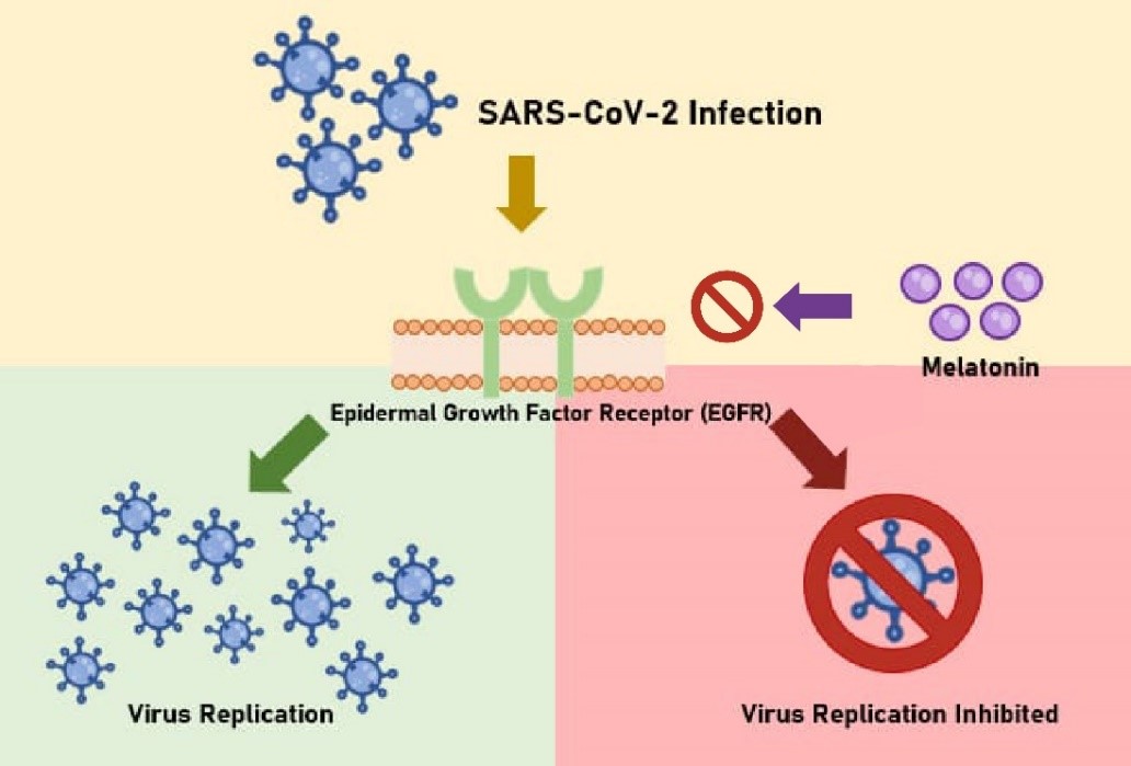
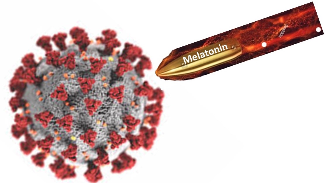
Vol 3 No 4 (2020)In this issue, we continue to report developments regarding the potentially therapeutic effects of melatonin on COVID-19. Based on the recent data that growth factor signaling is required for SARS-CoV-2 replication in the infected cells and the previous publications on the effects of melatonin on epidermal growth factor signaling, in this issue, Tesarik speculates that melatonin also acts against the virus itself via targeting the same molecular pathway to inhibit viral replication (see the cover figure). Ahmed suggests that COVID-19 patients should avoid room light during the night to preserve their endogenous melatonin production. This simple procedure may improve the immunity of COVID-19 patients and impact their outcomes. Thus, in the systemic review of melatonin and COVID-19, Pandi-Perumal and colleagues conclude that low melatonin may be a contributor to SARS-CoV-2 disease. Due to the still lack of a specifically effective treatment, any reasonable suggestion, idea or hypothesis that may improve the therapeutic outcome from this devastatingly infectious disease is helpful. In this regard, melatonin as an anti-inflammatory and antioxidant agent along with its large safety margin, low cost and ease of administration via multiple routes makes it an attractive molecule for this purpose. This issue also includes several other very interesting research articles and reviews which are related to different aspects of melatonin physiology.
-
 Melatonin and COVID-19 Special Issue
Vol 3 No 3 (2020)
Melatonin and COVID-19 Special Issue
Vol 3 No 3 (2020)Since the outbreak of COVID-19 in December 2019, several hundred thousand of infected individuals have lost their life and the global economy has been negatively impacted. Scientists and physicians have spent multiple resources to develop effective vaccines in an attempt to finally conquer this deadly pandemic so societies and the economies can return to normal. During the window period prior to the availability of an effective vaccine or the identification of reliable remedies to reduce the morbidity and mortality associated with COVID-19, scientists and clinicians have been confronted with dauting task of saving lives without known treatments. The society of melatonin scientists feel an obligation to contribute our knowledge and experience with others to combat this pandemic. This is the purpose for publishing this special issue entitled “Melatonin and COVID-19”. In this special issue, we have reviewed as many aspects of melatonin and COVID-19 as possible. Included in this series are reports of how the CNS, heart, lungs, gut and erythrocytes are affected by COVID-19 and what is the potential of melatonin on them. For the molecular mechanisms and signal transduction pathways, these involve effects of melatonin on CD147 binding with SARS-CoV-2 spike protein, the switch of aerobic glycolysis to mitochondrial oxidative phosphorylation in immune cells, ER stress-UPR-autophagy alterations and kynurenine and the alpha 7 nicotinic receptor activation. In addition, the melatonin doses that can be used in COVID-19 treatment have also be estimated based on the published reports. Most importantly, in this special issue, we have collected the first clinical report using melatonin to treat COVID-19 patients. Castillo et al. have successfully treated 10 patients with high dose of melatonin. Eight of these 10 patients were over 60 years old and 7 of them had the comorbidities of hypertension, diabetes and/or COPD. Advanced age and predisposed medical conditions are huge risk factors for mortality in COVID-19 patients. Fortunately, under melatonin treatment, all of the patients survived. This preliminary and important clinical data encourage further and large-scale clinical trials. We hope that this special issue will stimulate the enthusiasm for melatonin research in the field of deadly viral infections, particularly in relation to COVID-19 disease.
-
 Melatonin Research
Vol 3 No 2 (2020)
Melatonin Research
Vol 3 No 2 (2020)Melatonin membrane receptors have been extensively studied and well documented in animals. These receptors are classified into the high affinity MT1, MT2 and low affinity MT3 subtypes. Whether MT3 is a melatonin membrane receptor is still debatable since the best evidence shows that MT3 is situated in the cytoplasm rather than in the cell membrane. MT3 actually is a cytosolic enzyme, human quinone reductase 2 (hQN2). MT1 and MT2 are classic G protein coupled receptors and their activations often involve the cAMP pathway. Compare to animals, melatonin receptors in plants are a novel issue and many uncertainties exist. In 2018, Wei et al [Wei J, Li D, Zhang J, Shan C, Rengel Z, Song Z, Chen Qi (2018) Phytomelatonin receptor PMTR1-mediated signaling regulates stomatal closure in Arabidopsis thaliana, J. Pineal Res. 265 :e12500] reported a plant melatonin receptor which was referred as the first phytomelatonin receptor (CAND2/PMTR1) to be identified. Similar to the animal membrane receptors, they report that PMTR1 also couples with heterotrimeric G protein α subunit and regulates stomatal closure via H2O2 production and Ca2+ influx in Arabidopsis. However, this PMTR1 is challenged by the report published in this issue. Using state of the art technologies, Lee and Back show that CAND2/ PMTR1 protein localizes in the cytoplasm rather than in the plasma membrane in plants. In cand2 knockout mutant plants, melatonin-mediated mitogen-activated protein kinase (MAPK) activation was not abolished nor did melatonin-mediated defense gene inductions diminish compared to that in the wild type. The authors claim that PMTR1 is neither a melatonin membrane receptor, nor is it involved in the melatonin-mediated defense signaling pathway via G protein components. This important observation by Lee and Back raises a new question whether plants have the melatonin membrane receptors as do animals, and if they do, are they G protein coupled? The MT1 and MT2 in animals were acquired during evolution. In many ways, animals and plants have different evolutionary processes.
-
 Melatonin Research
Vol 3 No 1 (2020)
Melatonin Research
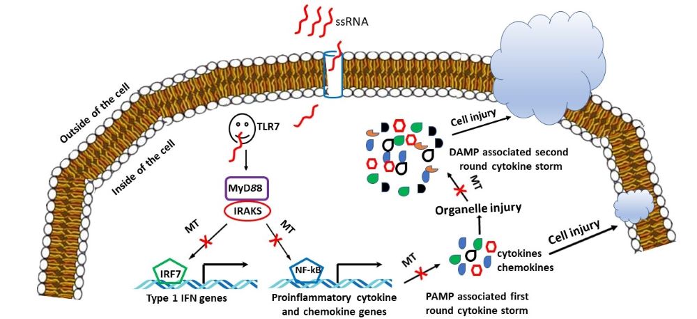
Vol 3 No 1 (2020)In this issue, the focus is on the potential utility of melatonin use for the unpredictable and rapidly-developing public health events such as in exposure to vesicant chemical warfare agents (Romero et al.), outbreak of Ebola infection (Reiter et al.) and the current COVID-19 pandemic (Tan & Hardeland). Unfortunately, as usual there is a lack of immediately available and effective treatments for these public health threats, thus, leading to high mortality. The underlining mechanisms for the high mortality commonly involves free radical attack and destructive inflammation in numerous tissues and organs. For example, the targeted tissue for the Ebola is vascular system while the lung is the target for sulfur mustards and COVID-19. In COVID-19, the free radical formation and massive inflammatory reaction is caused by the exaggerated innate immune response of the host against the pathogens. This hyperactive innate immune response is associated with the release of a large quantity of cytokines referred as a “cytokine storm”. This phenomenon is also observed in the vesicant chemical warfare agent exposure and in Ebola virus infection. It appears that the pathogens per se serve as the pathogen associated molecular patterns (PAMPs) triggering the primary “cytokine storm” and the damaged molecules of the host cell resulting from the primary “cytokine storm” function as damage associated molecular patterns (DAMPs) to cause the secondary “cytokine storm”. This becomes a vicious cycle; if this vicious cycle is not interrupted, it causes massive tissue and organ damage and sometimes death. All of the authors mentioned above believe that melatonin is a molecule which has the capacity to break this vicious cycle. Melatonin is a powerful free radical scavenger to reduce the tissue oxidative damage and it is also an effective anti-inflammatory agent to depress the “cytokine storm”. As a result, melatonin may increase the tolerance of the host to the pathogens and save precious time for the patients to develop an adaptive immune-response and finally recover from the pathogens’ attack. In addition, melatonin also promotes the adaptive immune response by increasing T lymphocyte proliferation and B cells to generate specific antibodies. Melatonin has a huge safety margin and is readily available. Based on its pharmacological and physiological characteristics, melatonin may exhibit beneficial effects if it is used in these unpredictable public health events including vesicant chemical warfare agents, Ebola and COVID-19 infections.
-
 Melatonin Research
Vol 2 No 4 (2019)
Melatonin Research
Vol 2 No 4 (2019)During evolution, almost all organisms that inhabited the earth had to adapted to the natural light/dark cycle to adjust their internal bio-circadian rhythms. However, these bio-circadian rhythms are disturbed by the increased global light pollution. The adverse consequences of light pollution are obvious in organisms, including on the health of humans. It is speculated that many human diseases including diabetes, obese, hypertension, neurodegenerative diseases and cancer are more or less associated with excessive nighttime light exposure. As a result, light at night has been classified as a Group 2A carcinogen by the International Agency for Cancer Research (IACR). The impact of light at night on the reproductive physiology of free living organisms has not been fully investigated. A few studies have observed the adverse effects of light pollution on the reproductive activities of birds. To address this issue in mammals, Berbets et al. exposed pregnant rats to constant light during their entire gestation period. The results were remarkable. None of the 14 pregnant dames gave birth. In contrast, all 10 dames under the normal light/dark cycle successfully delivered healthy pups. The morphological evidence showed that the pregnancy was terminated at an early stage of gestation. The unsuccessful pregnancy was accompanied by pineal gland dysfunction, low serum melatonin, and a uterine and systemic inflammation (see the graphical abstract). Such a profound effect of light pollution on mammalian reproduction has not been reported previously and it was unexpected. It is known that pinealectomy, which also eliminates the melatonin rhythm, impacts mammalian reproduction but not to this degree. Based on these observations, we probably have underestimated the influence of light at night on the gradually-increasing human infertility as observed in developed countries. The report is published in this issue of the journal (Berbets, A.M., Barbe, A.M. and Yuzko, O.M. 2019. Constant light exposure terminates pregnancy in rats with pineal gland dysfunction, low melatonin level and pro-inflammatory response. Melatonin Research. 2: 9-24. doi.10.32794/mr11250038). This observation requires confirmation in other species.
-
 Melatonin Research
Vol 2 No 3 (2019)
Melatonin Research
Vol 2 No 3 (2019)In this issue, Reiter and colleagues propose that melatonin in mitochondria, either synthesized de novo in this organelle or taken up from the circulation, is instrumental in reprogramming cancer cell glucose metabolism from the cytosol (the Warburg effect) to the mitochondria. Melatonin predictably achieves this action by inhibiting pyruvate dehydrogenase kinase (PDK) allowing for the upregulation of pyruvate dehydrogenase complex, which catalyzes the conversion of pyruvate to acetyl coenzyme A (acetyl CoA). Acetyl CoA feeds the citric acid cycle and increases ATP production via oxidation phosphorylation. Acetyl CoA also is required for the rate-limiting enzyme in melatonin production, arylalkylamine N-acetyltransferase (AANAT), which is in mitochondria to convert serotonin to N-acetylserotonin. This ensures melatonin production in the mitochondria of both normal cells and cancer cells. Cytosolic glycolysis is characteristic of many solid tumor cells and enhances their ability to rapidly proliferate, to avoid apoptosis and to become metastatic. By redirecting glucose metabolism from the cytosol to the mitochondria in cancer cells, melatonin reduces tumor cell growth, subjects them to higher levels of oxidative stress and increases apoptotic cell death. Reiter and colleagues speculate that this may be an essential mechanism by which melatonin inhibits Warburg-dependent cancer growth. This novel hypothesis may be a major step in understanding how melatonin limits tumor growth. The hypothesis is easily testable and this will likely happen quickly.
-
 Melatonin research
Vol 2 No 2 (2019)
Melatonin research
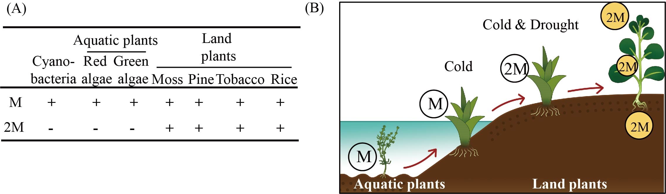
Vol 2 No 2 (2019)In this issue, Lee and Back publish an article (Lee, H.-J. and Back, K. 2019. 2-Hydroxymelatonin confers tolerance against combined cold and drought stress in tobacco, tomato, and cucumber as a potent anti-stress compound in the evolution of land plants. Melatonin Research. 2, 2, 36-47. DOI:https://doi.org/https://doi.org/10.32794/mr11250020.) in which the authors identified 2-hydroxymelatonin as the major metabolite of melatonin. 2-Hydroxymelatonin is formed from melatonin as a result of the enzymatic activity of melatonin 2-hydroxylase. This enzyme is absent in aquatic plants including cyanobacteria and alga and is present only in land plants. 2-Hydroxymelatonin not only exhibits much higher levels than that of melatonin in plants but it also protects land plants from the combined stressors such as cold and drought.
Based on the observations, the authors propose a very interesting hypothesis. When the aquatic plants transformed into land plants, they faced the common challenges of cold and drought which rarely occur in aquatic environments. Under these combined stressors, melatonin per se could not provide sufficient protection as the data indicate. Thus, the land plants evolved melatonin 2-hydroxylase to generate 2-hydroxymelatonin to overcome the stressors of their terrestrial environments.
The hydroxylmelatonins have also been found in animals; however, they are speculated to be the products of melatonin’s interactions with reactive oxygen species (ROS) and reactive nitrogen species (RNS). Melatonin hydroxylases have not yet been identified yet in animals. The potential functions of hydroxylmelatonins and their synthetic pathways in animals would be interesting topics of exploration.
-
 Melatonin research
Vol 2 No 1 (2019)
Melatonin research
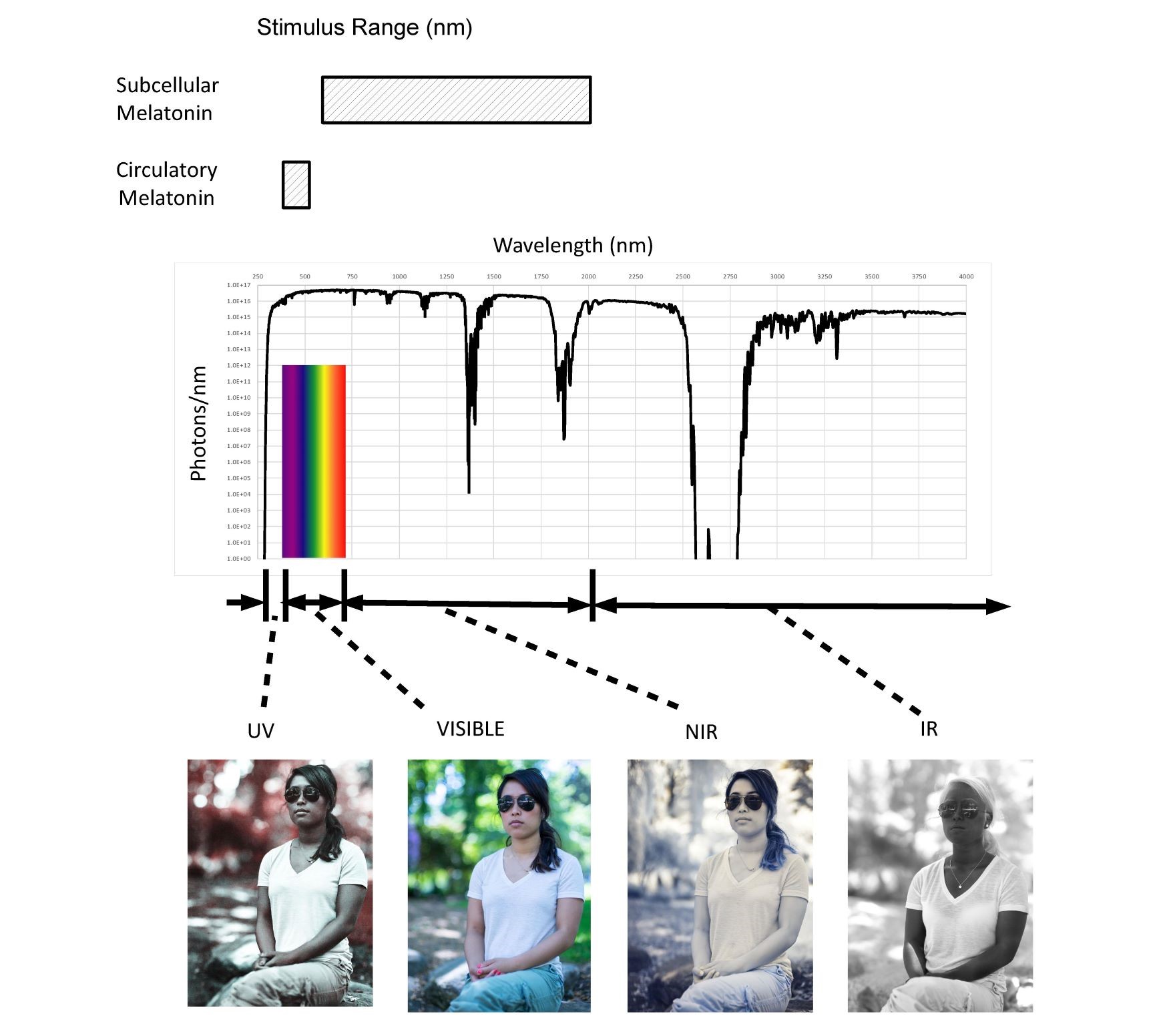
Vol 2 No 1 (2019)In the current issue, S. Zimmerman and RJ. Reiter published an article entitled “Melatonin and the Optics of the Human Body”. In this article they pointed out that the active wavelengths for inhibition of circulatory melatonin extend from 420nm to 500nm (visible). Optically they show that uniquely virtually all our cells are exposed to near infrared (NIR) photons which extend from 650nm to 1200nm. NIR represents 70% of the total solar spectrum from a photochemical viewpoint (photons/second). While this evidence supports that the visible light suppresses the circulatory melatonin produced by the pineal gland, the literature and bio-optical models appear to support the hypothesis that the NIR portion of natural sunlight stimulates subcellular melatonin synthesis in large amounts in most of our cells throughout the day. They hypothesized that the role of circulatory melatonin produced by the pineal gland is to provide an efficient method of delivering supplemental melatonin during periods of low cellular activity, while the subcellular melatonin generated from solar NIR stimulation forms an antioxidant reservoir protecting damaged or aging cells in both diurnal and nocturnal animals. They mentioned that circulatory melatonin may be the “Hormone of Darkness”, subcellular melatonin may be the “Hormone of Daylight”. This figure illustrates the broad range of optical properties that the human body is exposed to throughout the solar spectral range from 250nm to over 4000nm and their association with melatonin synthesis.
-
 Inaugural Issue of Melatonin Research
Vol 1 No 1 (2018)
Inaugural Issue of Melatonin Research
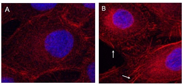
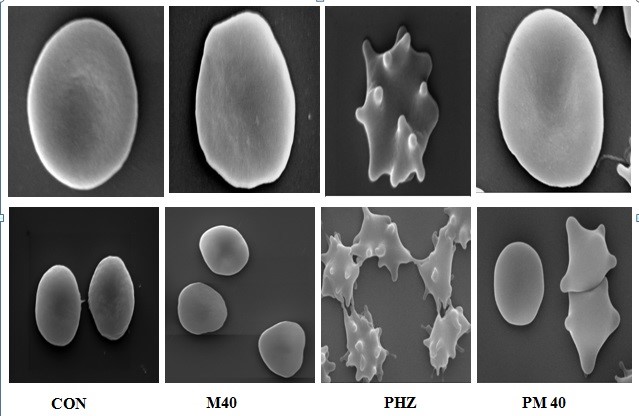
Vol 1 No 1 (2018)Phenylhydrazine (PHZ) treatment induces the red blood cell (RBC) phenotype changes resembling the severe β-thalassaemia including the rigidly and mechanically unstable membranes of RBC in conjunction with the oxidized alpha-globin chains which further connect to the membrane skeleton. These alterations are related to the RBC oxidative stress and iron-overloading. Melatonin as a potent antioxidant effectively prevents RBC deformation caused by PHZ treatment. The result suggest that melatonin may have the therapeutic potential to β-thalassaemia in particular and also to other hemolytic anemias involving oxidative stress in general.
CON: control; PHZ: phenylhydrazine; M40: melatonin (40 nmoles/ml); PM40: PHZ plus melatonin (40 nmoles/ml)
1 - 23 of 23 items

